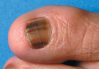
Melanoma occurs when the skin’s pigment-producing cells called melanocytes become malignant.
Clusters of melanocytes can also produce non-cancerous growths called moles. When it first develops, melanoma may appear as a new mole, but it quickly distinguishes itself from a normal mole in shape, color, and texture.
Acral Lentiginous Melanoma


One of best ways to catch melanoma in its early stage is to watch for moles that look abnormal.
Melanoma is:
- Is one of the most common cancers
- Can develop on any skin surface, including areas of skin completely sheltered from the sun and under the nails.
- Causes the greatest number of skin cancer–related deaths worldwide
- Occurs most frequently in fair-skinned people
Learning the ABCDE’s help patient to know what to look for. Early detection is key.
These rules are at their greatest diagnostic accuracy when used in combination:
- Asymmetry: Half the lesion does not match the other
- Border irregularity: edges are ragged, notched, or blurred
- Color variegation: pigmentation is not uniform and may display shades of tan, brown, or black, white, reddish, or blue discoloration
- Diameter: diameter greater than 6 mm is characteristic, although some melanomas may have smaller diameters
- Evolving: Changes in lesion over time are characteristic, critical for nodular or amelanotic (unpigmented) melanoma
Acral Lentiginous
An uncommon form of the cancer melanoma which can occur under the nail plate or at the ends of the fingers or toes is called acral lentiginous melanoma.
When occurring under the nail it can easily be mistaken for a fungal infection or harmless streak of pigment or dried blood however if not diagnosed early it has the same potential serious consequences as the other forms of melanoma.
Any discoloration of the nail or distant parts of the fingers or toes persisting more than a few weeks should be evaluated by a dermatologist as soon as possible regardless of what the suspected cause of the pigmentation is thought to be.
Am I At Risk?
Risk factors involve certain physical characteristics: blue/green eyes, blond or red hair, and a light complexion.
Other factors that increase a person’s risk for developing melanoma are sun sensitivity, an occurrence of blistering sunburn(s) in childhood and adolescence, weakened immune system, personal and family history of melanoma or skin cancer, and regular exposure to UV radiation.
What Are the Treatment Options?
The process of selecting a treatment will be decided upon after biopsy and during staging.
For melanoma that is in its early growth phase and Stage I, can be removed surgically by Dr. Michalak in the office. The tumor as well as some healthy tissue around it and generally the wound will be closed with stitches afterward.
For more invasive and advanced melanoma studies of the lymph glands may need to be performed prior to the removal of the melanoma surgically and may be followed by chemotherapy.
The ABCDEs of Melanoma Detection
Look for danger signs in pigmented lesions of the skin. Consult your dermatologist immediately if any of your moles or pigmented spots exhibit:
 |
One half unlike the other half. |
 |
Irregular, scalloped or poorly defined border. |
 |
Varied from one area to another; shades of tan and brown, black; sometimes white, red or blue. |
 |
While melanomas are usually greater than 6mm (the size of a pencil eraser) when diagnosed, they can be smaller. |
 |
A mole or skin lesion that looks different from the rest or is changing in size, shape or color. |
We encourage you to contact us if you have a family history of melanoma, have any spots that look like the pictures or are very dark, bleed and does not heal. Appointments can be schedule by calling 425-391-2500.
